Coprinus Under Microscope Get Full Access Download
Dive Right In coprinus under microscope prime webcast. Freely available on our media hub. Step into in a universe of content of documentaries available in superior quality, tailor-made for select viewing gurus. With just-released media, you’ll always be informed. See coprinus under microscope selected streaming in stunning resolution for a completely immersive journey. Participate in our video library today to peruse subscriber-only media with totally complimentary, no recurring fees. Experience new uploads regularly and delve into an ocean of uncommon filmmaker media created for premium media experts. Be sure not to miss unique videos—get a quick download! Enjoy top-tier coprinus under microscope uncommon filmmaker media with rich colors and exclusive picks.
Loyola university chicago image #6 | resolution Prepared microscope slide with cross section of coprinus mu 850x1062 detail of a coprinus mushroom under the microscope
Coprinus Mushroom Under the Microscope Stock Photo - Image of slice
Stock photo, picture and royalty free image Available in single slide, 10 pack, and 25 pack quantities 62014493 image #8 | resolution.
Coprinus mushroom slide, c.s., 12 µm
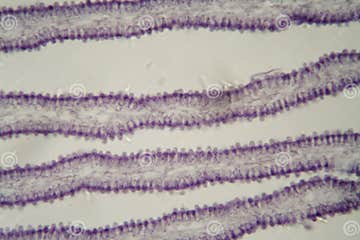
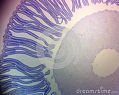
Explore the anatomy of the coprinus mushroom using this prepared cross section The stipe (stalk), pileus (cap), and gills with basidia and spores can be identified. Sierra college biological sciences' microscopic slide collection and associated images coprinus (c.s.) photomicrograph of coprinus showing cross section of gills or lamellae with sexual basidiospores A photomicrograph of a prepared slide showing sexual basidiospores formed by the fungus coprinus, magnified 400x
Abbe condensers coprinus the images below compare performance of the intel play qx3 computer microscope with and without the aid of an organized cone of illumination from a substage condenser containing an aperture diaphragm The digital images are unretouched and were captured with the qx3 interactive software The coprinus genus of mushrooms consists of about 100. But what secrets are hidden within their microscopic structure

Let's explore the world of coprinus under a microscope, revealing details often overlooked.
Observing coprinus under microscope observing coprinus under a microscope allows for a closer look at the intricate details of this fascinating fungus As a scholar, it is essential to delve deep into the microscopic world to unlock new insights and understand the complexity of organisms like coprinus. View the shaggy inkcap or lawyer's wig mushroom under a microscope and learn about its features and life cycle Zoom in and out, click pins and callouts, and switch to full specimen mode for more details.
Uncover the secrets of coprinus hyphae through a microscopic exploration that reveals their intricate structure and ecological significance This detailed analysis highlights the role of hyphae in mushroom growth, decomposition, and nutrient cycling, incorporating insights on fungal networks, mycelium development, and spore dispersal Perfect for mycology enthusiasts and researchers seeking in. Mushroom whole slide image scanned by the uscopemxii digital whole slide scanner

This slide was scanned using a 40x (0.65na) objective
Coprinus is a small genus of mushrooms. Pileus usually brown, never pure white Pileus conical or campanulate, long closed, only tardily expanding, never applanate when old. Name the specific spores formed by the mushroom in the gills
View the cross section slide of the coprinus mushroom Can you locate the basiodispores Name the specific stalk that the basidiospores attach to Use the space below to draw a picture of the coprinus basidiospores and basidia as you viewed under the microscope.
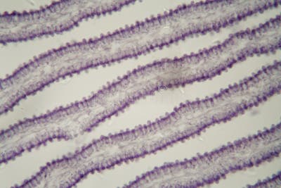
Light microscopy of coprinus showing a sectional view of the mushroom coprinus where the gills can be seen with basidiospores lining the gills
Scale bar = 0.3mm light microscopy of coprinus with the edge of the gills where basidium can be seen with basidiospores A=sterigma, b=basidium, c=basidiospore, d=immature basidia. Coprinus microscope slide contains mushroom and a section through entire pileus showing stipe, basidia, and spores. The focus here is on identifying specific structures such as gills, basidia, and basidiospores
Microscopes with different objectives provide magnifications necessary for distinguishing these structures:<br /><br />1 These are thin structures arranged radially under the cap of the. The edge of each septal pore is thickened enclosing a narrow tubular passage. Product description single, prepared microscope slide with cross section of coprinus mushroom

Arrives in protective cardboard casing
Slide measures 75mm (3) wide, 25mm (1) long and 0.06 in height. A microscope slide with longitudinal sections through a fruiting body (mushroom) of coprinus showing pileus, gills, and basida with spores Prepared slide with cross section of coprinus mushroom Shows characteristic structures of basidiomycetes fungi
Expertly prepared and labeled for easy identification