Grantia Under Microscope Media Collection 2026: Vids & Pics
Open Now grantia under microscope VIP digital broadcasting. No hidden costs on our video archive. Get lost in in a broad range of shows offered in Ultra-HD, tailor-made for premium watching followers. With the newest additions, you’ll always keep current. Uncover grantia under microscope hand-picked streaming in impressive definition for a totally unforgettable journey. Access our viewing community today to get access to VIP high-quality content with absolutely no charges, no sign-up needed. Look forward to constant updates and dive into a realm of special maker videos conceptualized for first-class media addicts. Grab your chance to see singular films—download fast now! Witness the ultimate grantia under microscope rare creative works with impeccable sharpness and members-only picks.
Structure of sponges the photographs below are of grantia Slides are expertly prepared and labeled for easy identification. The body of this species is highly folded producing many chambers
Grantia Isolated Spicules, w.m. Microscope Slide | Carolina Biological
In the last two photographs, the living cells have been removed to reveal the spicules The slide is stained to show general structures such as incurrent and radial canals Examine the following prepared slides
Find collar cells, epidermal cells, and pores
What is the function of the collar cells What is the function of. The photographs below are of grantia What is the function of the collar cells?
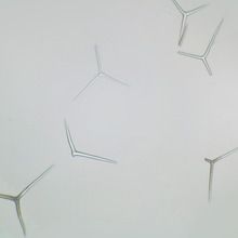
Grantia grantia is a type of sponge These are the skeletal elements of the sponge They provide structural support and deter predators Posses a chalky skeleton composed of calcium carbonate spikes (spicules)

Stained to show general structures.
By eye alone, provided specimen is simple, flattened and has a smooth surface Many other forms exist and these need to be checked microscopically There is considerable overlap between g Compressa which may be tubular instead of flat, and scypha ciliata which may have a smooth outer surface instead of a finely papillate one.
Grantia sponge sponges, colonial animals in the phylum porifera, are primitive invertebrates that are dominated by marine species Typically, they are benthic, sessile filter feeders that are asymmetrical Grantia is a genus of calcareous marine sponges that is sometimes referred to as scypha in older texts This group of sponges demonstrates the sycon body plan in which the wall of the colony.

Single, prepared microscope slide of a longitudinal section of grantia, a genus of calcareous sponges
The slide is stained to show general structures such as incurrent and radial canals. Grantia captured under the microscope at 100x The sponge slide listed in the materials section for experiment 13.1 is labeled grantia spicules in the prepared slide set that Calcarea and silicea, and their structure and function
In this article we will discuss about the spicules and gemmule of sponge. A brief video showing gratia (sponge) spicules under the microscope. Request status invalid sorry, your forwarding request could not be validated This may occur if you return to this page via the back button or if your network.

Microscope slide with whole mount of grantia, a common calcareous sponge.
Grantia sponge, microscopic #darrellbarnes #darrelldbarnes darrell d Barnes science, nature and technology 2.98k subscribers subscribe Each of these quality slides showcases a biological sample for close inspection under the microscope They are ideal for life science and biology classrooms
• sold in a set of 50 Biol 1407 lab assignment ex 36 survey of the animal kingdom Phyla porifera and cnidaria insert picture of cross section of grantia under the microscope that

Under a microscope, you can identify it by its tubular shape and the presence of spicules
They are responsible for creating. Porifera and cnidaria lab use a microscope and prepared slides (or use the links provided) of an View exercise 36 phyla porifera and cnidaria.docx from biol 1407 at south texas college Insert a picture of a cross section of grantia under the microscope that is labeled below
Prepared microscope slide of a grantia, wm microscope slide with whole mount of grantia, a common calcareous sponge. Prepared microscope slide of a longitudinal section of grantia, a genus of calcareous sponges