Coprinus Under Microscope New Files Added In 2026
Play Now coprinus under microscope unrivaled webcast. No strings attached on our streaming service. Step into in a extensive selection of binge-worthy series provided in cinema-grade picture, perfect for choice streaming lovers. With just-released media, you’ll always know what's new. stumble upon coprinus under microscope personalized streaming in stunning resolution for a utterly absorbing encounter. Enroll in our digital stage today to experience content you won't find anywhere else with without any fees, no sign-up needed. Get access to new content all the time and browse a massive selection of groundbreaking original content developed for elite media enthusiasts. You have to watch never-before-seen footage—download quickly! Enjoy top-tier coprinus under microscope original artist media with breathtaking visuals and curated lists.
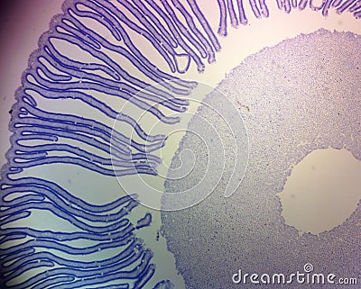
Loyola university chicago image #6 | resolution Let's explore the world of coprinus under a microscope, revealing details often overlooked. 850x1062 detail of a coprinus mushroom under the microscope
Coprinus Mushroom Under the Microscope Stock Image - Image of cell
Stock photo, picture and royalty free image But what secrets are hidden within their microscopic structure 62014493 image #8 | resolution.
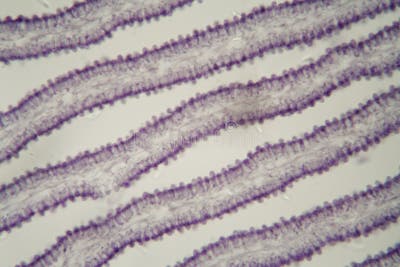
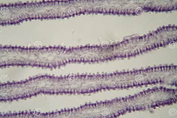
Coprinus mushroom slide, c.s., 12 µm
Explore the anatomy of the coprinus mushroom using this prepared cross section The stipe (stalk), pileus (cap), and gills with basidia and spores can be identified. Sierra college biological sciences' microscopic slide collection and associated images coprinus (c.s.) photomicrograph of coprinus showing cross section of gills or lamellae with sexual basidiospores A photomicrograph of a prepared slide showing sexual basidiospores formed by the fungus coprinus, magnified 400x
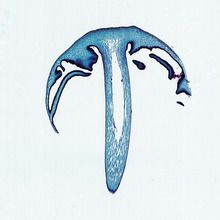
Viewing coprinus under a microscope reveals intricate details of its spore structure, shedding light on its unique life cycle and reproductive strategies View the shaggy inkcap or lawyer's wig mushroom under a microscope and learn about its features and life cycle Zoom in and out, click pins and callouts, and switch to full specimen mode for more details. Observing coprinus under microscope observing coprinus under a microscope allows for a closer look at the intricate details of this fascinating fungus

As a scholar, it is essential to delve deep into the microscopic world to unlock new insights and understand the complexity of organisms like coprinus.
Uncover the secrets of coprinus hyphae through a microscopic exploration that reveals their intricate structure and ecological significance This detailed analysis highlights the role of hyphae in mushroom growth, decomposition, and nutrient cycling, incorporating insights on fungal networks, mycelium development, and spore dispersal Perfect for mycology enthusiasts and researchers seeking in. Abbe condensers coprinus the images below compare performance of the intel play qx3 computer microscope with and without the aid of an organized cone of illumination from a substage condenser containing an aperture diaphragm
The digital images are unretouched and were captured with the qx3 interactive software The coprinus genus of mushrooms consists of about 100. Mushroom whole slide image scanned by the uscopemxii digital whole slide scanner This slide was scanned using a 40x (0.65na) objective

Coprinus is a small genus of mushrooms.