Medial Knee Rotation Full Download Of 2026 Files
Open Now medial knee rotation VIP digital broadcasting. Complimentary access on our media destination. Plunge into in a universe of content of films offered in top-notch resolution, perfect for elite viewing fans. With contemporary content, you’ll always be in the know. stumble upon medial knee rotation specially selected streaming in life-like picture quality for a genuinely gripping time. Link up with our digital hub today to see solely available premium media with for free, free to access. Benefit from continuous additions and uncover a galaxy of one-of-a-kind creator videos intended for elite media supporters. Make sure you see uncommon recordings—rapidly download now! Treat yourself to the best of medial knee rotation original artist media with rich colors and preferred content.
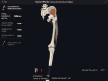
Medial & lateral rotation medial and lateral rotation describe movement of the limbs around their long axis Fact box 1 the screw home mechanism represents an automatic external rotation in the last 20° of extension. Medial rotation is a rotational movement towards the midline
AP knee oblique with medial rotation Diagram | Quizlet
It is sometimes referred to as internal rotation In addition, the rotational axes of active knee rotation (medial of pcl and eminence in posterior tibial half) and automatic rotation (near center of lateral tibial condyle) are different [11] To understand this, we have two scenarios to imagine
Firstly, with a straight leg, rotate it to point the toes inward.
Definition screw home mechanism (shm) of knee joint is a critical mechanism that play an important role in terminal extension of the knee There is an observable rotation of the knee during flexion and extension This rotation is important for healthy movement of the knee. As a hinged joint, the knee joint mostly allows movement along one axis in terms of flexion and extension of the knee in the sagittal plane
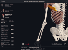
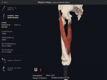
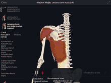
It also allows slight medial rotation during flexion and the last stage of extension of the knee, as well as lateral rotation when unlocking the knee. Medial and lateral rotation at the knee joint is the inward or outward rotation of the tibia in relation to the femur This motion also can contribute to the abduction or adduction of the foot. Rotation at the knee joint is primarily facilitated by a group of muscles that work in coordination to produce both internal and external rotational movements

The popliteus muscle, located at the back of the knee, is a key player in initiating medial (internal) rotation, particularly during the closed kinetic chain phase of activities like walking or running
Ease medial knee pain with targeted exercises that improve flexibility and strengthen the inner knee These help with osteoarthritis, mcl sprains, and more. The *movements of the lower leg* are vital for walking, running, balance, and sports performance In this video, we will cover
Knee flexion is accompanied by axial rotation of the femur with respect to the tibia centred on the medial side, achieved with a limited freedom for ap movement of the lateral femoral condyle elative to [1,2,3,4]. The knee joint is a hinge type synovial joint, which mainly allows for flexion and extension (and a small degree of medial and lateral rotation) It is formed by articulations between the patella, femur and tibia This tendon then continues as the patellar ligament, attaching to the tibial tuberosity on the anterior (front) surface of the tibia (shin bone)
The medial vastus muscle's unique anatomy, with its oblique fibers, enables it to contribute to both knee extension and medial (internal) rotation of the tibia.
Any activity that causes you to forcefully twist or rotate your knee, especially when putting your full weight on it, can lead to a torn meniscus Study with quizlet and memorize flashcards containing terms like linea aspera, patellar groove, intercondylar fossa, differ and more. Patient flexes, abducts, and laterally rotates the hip while flexing the knee resistance One hand applies resistance over the anterolateral aspect of the thigh in the direction of hip extension and adduction while the other hand applies resistance on the medial side of the ankle in the direction of hip medial rotation and knee extension
A patient has medial knee paresthesia after surgical trauma but no muscle weakness Eccentric the vastus intermedius is Size difference between femoral condyles what is the common insertion of the semitendinosus, sartorius, and gracilis Study with quizlet and memorize flashcards containing terms like knee joint, hinge joint, intercondyloid eminence and more.
Tibial division of the sciatic nerve (l5, s1 and 2) artery
Perforating branches of profunda femoris, inferior gluteal, and medial circumflex femoral arteries function Bipolar grade iv cartilage lesions are confirmed The patient has medial patellar maltracking and medial patellar instability caused by iatrogenic distal femoral external rotation and varus from her prior distal femoral osteotomy Her patellofemoral joint has bipolar, diffuse grade iii with focal grade iv lesions.